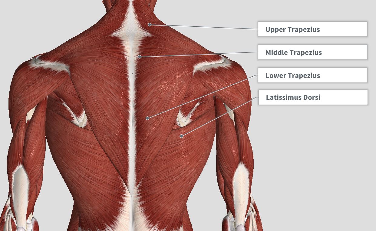
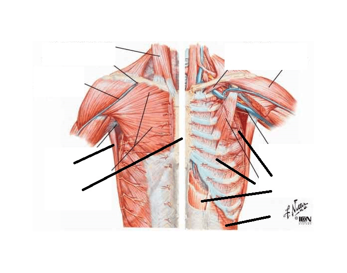
Anterior View of Thoracic Skeletal Structures 6 Biology Diagrams The thorax is divided into bony structures, muscles, vessels, nerves, and the thoracic cavity.Below is a detailed breakdown of its anatomy: Boundaries of the Thorax. The thorax has defined anatomical boundaries: Superior Boundary: Thoracic inlet (or superior thoracic aperture) at the base of the neck.; Inferior Boundary: Diaphragm, separating the thoracic cavity from the abdominal cavity. The thorax forms from the thoracic wall, its superficial structures (breast, muscles, and skin), and the thoracic cavity. A thorough comprehension of the anatomy and function of the thorax will help identify, differentiate, and treat the plethora of pathology that can occur within the thorax. Thoracic wall The first step in understanding thorax anatomy is to find out its boundaries. The thoracic, or chest wall, consists of a skeletal framework, fascia, muscles, and neurovasculature - all connected together to form a strong and protective yet flexible cage.. The thorax has two major openings: the superior thoracic aperture found superiorly and the inferior thoracic aperture

The thoracic cage is made up of: (1) 12 thoracic vertebrae (see Chapter 3 ); (2) 12 ribs and their costal cartilages and (3) the sternum. THE RIBS. There are usually 12 pairs of ribs (i.e., the ribs that articulate with the 12 thoracic vertebrae from T1 to T12); however, there is a variation in rib number (see variant anatomy in the ribs).

Thorax: Anatomy, wall, cavity, organs & neurovasculature Biology Diagrams
The thorax (pl.: thoraces or thoraxes) [1] or chest is a part of the anatomy of mammals and other tetrapod animals located between the neck and the abdomen. [2] [3]In insects, crustaceans, and the extinct trilobites, the thorax is one of the three main divisions of the body, each in turn composed of multiple segments.. The human thorax includes the thoracic cavity and the thoracic wall. Anatomy. Section 1. Anatomy Thorax. Plate 1-1. Plate 1-2. Plate 1-3. Plate 1-4. Before describing the anatomy of the heart, it is helpful to review other anatomic features of the thoracic cavity and organs. (T1)—gives access to the root of the neck and is not closed by a specific structure. The thorax is the superior part of the trunk extending between the neck and the abdomen.It consists of several components:. Thoracic wall; Several cavities; Nerves, blood vessels, lymphatics; Internal organs; Breasts; Thoracic wall. The thoracic wall consists mainly of muscles and bones that form the thoracic cage.Overall, the thoracic wall is formed by the following structures:

The thoracic cavity is a central compartment within the upper part of the torso, enclosed by the ribs, the vertebral column, and the sternum.It houses vital organs such as the heart, lungs, and major blood vessels, as well as portions of the esophagus and trachea.. Location. The thoracic cavity is located in the chest region, extending from the base of the neck to the diaphragm, and is bounded

Thorax (overview) Biology Diagrams
The thorax is the area of the body situated between the neck and the abdomen. The thorax itself can be split up into various areas that contain important structures.. The thorax is bound by bony structures including the 12 pairs of ribs and thoracic vertebrae, whilst also being supported by many ligaments and muscles.. The muscles of the thorax are also important for the vital actions of Your thoracic cavity is a space in your chest that contains your heart, lungs and other organs and tissues. Overview Function Anatomy Conditions and Disorders Care. Contents. This layer of tissue helps protect the organs and structures inside your thoracic cavity. Conditions and Disorders.

