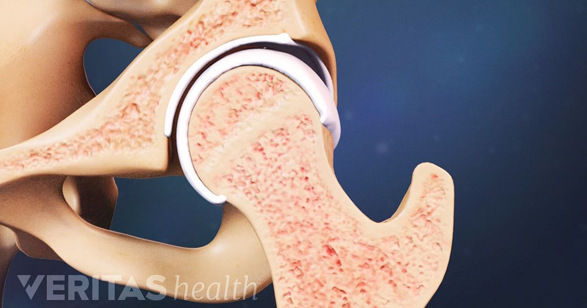
Bones Ligaments Quiz Biology Diagrams Hip ligament injury can be repaired, especially when they sustain significant damage or complete tears. Though not a ligament, the labrum is a ring of cartilage that goes around the outside rim of the socket of your hip joint. It acts like a gasket to hold the ball at the top of your thighbone securely within your hip socket. Athletes and The hip joint is the articulation between the ellipsoid head of the femur and the hemispherical concavity of the acetabulum located on the lateral aspect of the hip bone.The femoral head is covered with articular cartilage with the exception of a rough central depression, the fovea capitis, which is a surface of attachment for the ligament of the femoral head (ligamentum teres capitis femoris).
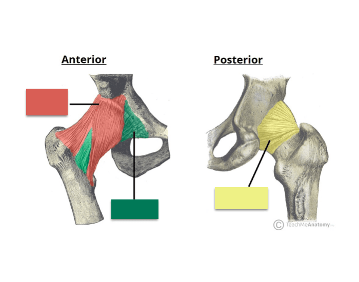
Your hip joint connects your thigh bone (femur) to your hip bone (pelvis). Your hip joint connects your thigh and hip bones. Cartilage: Cartilage is a smooth substance that covers the top of your thigh bone (femoral head) and the acetabulum socket. This substance is a cushion that absorbs impact when you walk and move. Ligaments are The pelvis is a girdle of bones, connected at the front by cartilage pad, called the pubis, and at the back by the lowest four fused vertebrae (the sacrum). The most notable ligaments in the hip joint are: Iliofemoral ligament, which connects the pelvis to the femur at the front of the joint. It keeps the hip from hyper-extension The hip joint is a ball-and-socket type joint and is formed where the thigh bone (femur) meets the pelvis. The femur has a ball-shaped head on its end that fits into a socket formed in the pelvis, called the acetabulum. Large ligaments, tendons, and muscles around the hip joint hold the bones (ball and socket) in place and keep it from dislocating.

Sports Medicine Review Biology Diagrams
The joint allows for flexion/extension, abduction/adduction, internal and external rotation. These actions are supported by a complex structure composed of muscles, tendons, ligaments, cartilage and neurovascular bundles. This post seeks to summarize, highlight and review key anatomy of the hip joint. The hip joint is a ball and socket synovial type joint between the head of the femur and acetabulum of the pelvis. It joins the lower limb to the pelvic girdle. Both the acetabulum and head of femur are covered in articular cartilage, Ligaments. The ligaments of the hip joint act to increase stability. They can be divided into two

Ischiofemoral ligament: It attaches to the posterior surface of the acetabular rim and labrum and courses circumferentially around the joint to its insertion on the anterior aspect of the femur.The ischiofemoral ligament limits internal rotation and hip adduction with flexion. Iliofemoral ligament (Y Ligament of Bigelow): It is a triangle-shaped ligament that attaches along the Ligaments, tendons, and muscles play an important role in the function of the hip. Ligaments are soft tissue structures that connect bones to bones.A joint capsule is a watertight sac that surrounds a joint.In the hip, the joint capsule is formed by a group of three strong ligaments that connect the femoral head to the acetabulum.