
Cell Cycle Graph Biology Diagrams The cell cycle is driven by precise temporal coordination among many molecular activities. To understand and explore this process, we developed the Cell Cycle Browser (CCB), an interactive web interface based on real-time reporter data collected in proliferating human cells. This tool facilitates vi … Regress out cell cycle scores during data scaling. We now attempt to subtract ('regress out') this source of heterogeneity from the data. For users of Seurat v1.4, this was implemented in RegressOut.However, as the results of this procedure are stored in the scaled data slot (therefore overwriting the output of ScaleData()), we now merge this functionality into the ScaleData() function itself.

The visualization of intricate cell cycle events such as DNA replication, chromatin condensation and contractile ring formation is a more robust approach to assess cell cycle progression. For example, fluorescently labelled PCNA can be used to track cell cycle progression . The distribution width of PCNA is narrow during the G1- and G2-phases The cell cycle is driven by precise temporal coordination among many molecular activities. To understand and explore this process, we developed the Cell Cycle Browser (CCB), an interactive web This visual essay introduces an advanced data-driven model and visualization tool that captures human cell division by integrating 3D data from 75 single-cell images. The model and the visualizations in the essay follow 15 cell structures through the whole cell cycle. Cell Structure Observations. Audience: advanced high school level and up

The Cell Cycle Browser: an interactive tool for visualizing, simulating ... Biology Diagrams
The left section of the CCB contains a network visualization of the cell cycle model (Figure 1-4) as well as controls for adjusting the model and simulation parameters (Figure 1-5). In a model, each molecular activity is referred to as a species —a pool of entities that are assumed to be indistinguishable, located in the same cellular In this issue, Sakaue-Sawano et al. (2008) present a method to visually distinguish cells at different phases of the cell cycle by the expression of colored fusion proteins that are under the control of the ubiquitin ligases SCF and APC. New imaging methods now permit the visualization of cellular events and phenomena in living cells and

Cell Systems Tool The Cell Cycle Browser: An Interactive Tool for Visualizing, Simulating, and Perturbing Cell-Cycle Progression David Borland,1,6 Hong Yi,1,6 Gavin D. Grant,2,3 Katarzyna M. Kedziora,4 Hui Xiao Chao,4,5 Rachel A. Haggerty,4,5 Jayashree Kumar, 5Samuel C. Wolff, 4Jeanette G. Cook,2,3 and Jeremy E. Purvis3 ,7 * 1Renaissance Computing Institute, University of North Carolina

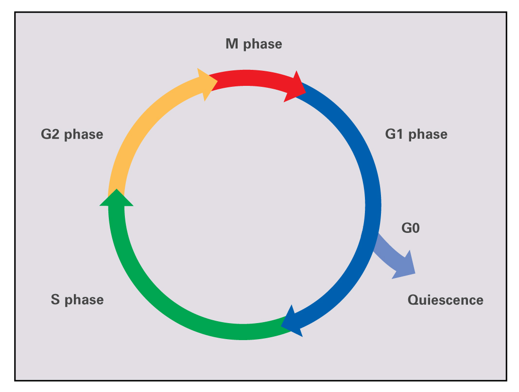
Cell Cycle Indicator Biology Diagrams
Cell Cycle Visualization Website Cesar Smokowski, Shuai Lin, Donghyeon Shin CS 4624 Multimedia, Hypertext, and Information Access, Dr. Fox Virginia Tech, Blacksburg VA 24061 Boldt, R. C. "The Cell Cycle - Phases - Mitosis - Regulation." TeachMePhysiology, TeachMe Science Series, 26 Jan. 2021,
FUCCI (Fluorescent Ubiquitination-based Cell Cycle Indicator) is a set of fluorescent probes which enables the visualization of cell cycle progression in living cells. FUCCI utilizes the phase-dependent nature of replication licensing factors Cdt1 and Geminin. A fusion protein of a fragment of Cdt1 (amino acids 30-120) with the fluorescent
