Draw A Cancer Cell Free Transparent Biology Diagrams The cell cycle is an evolutionarily conserved process necessary for mammalian cell growth and development. Because cell-cycle aberrations are a hallmark of cancer, this process has been the target of anti-cancer therapeutics for decades. However, despite numerous clinical trials, cell-cycle-targeting agents have generally failed in the clinic.
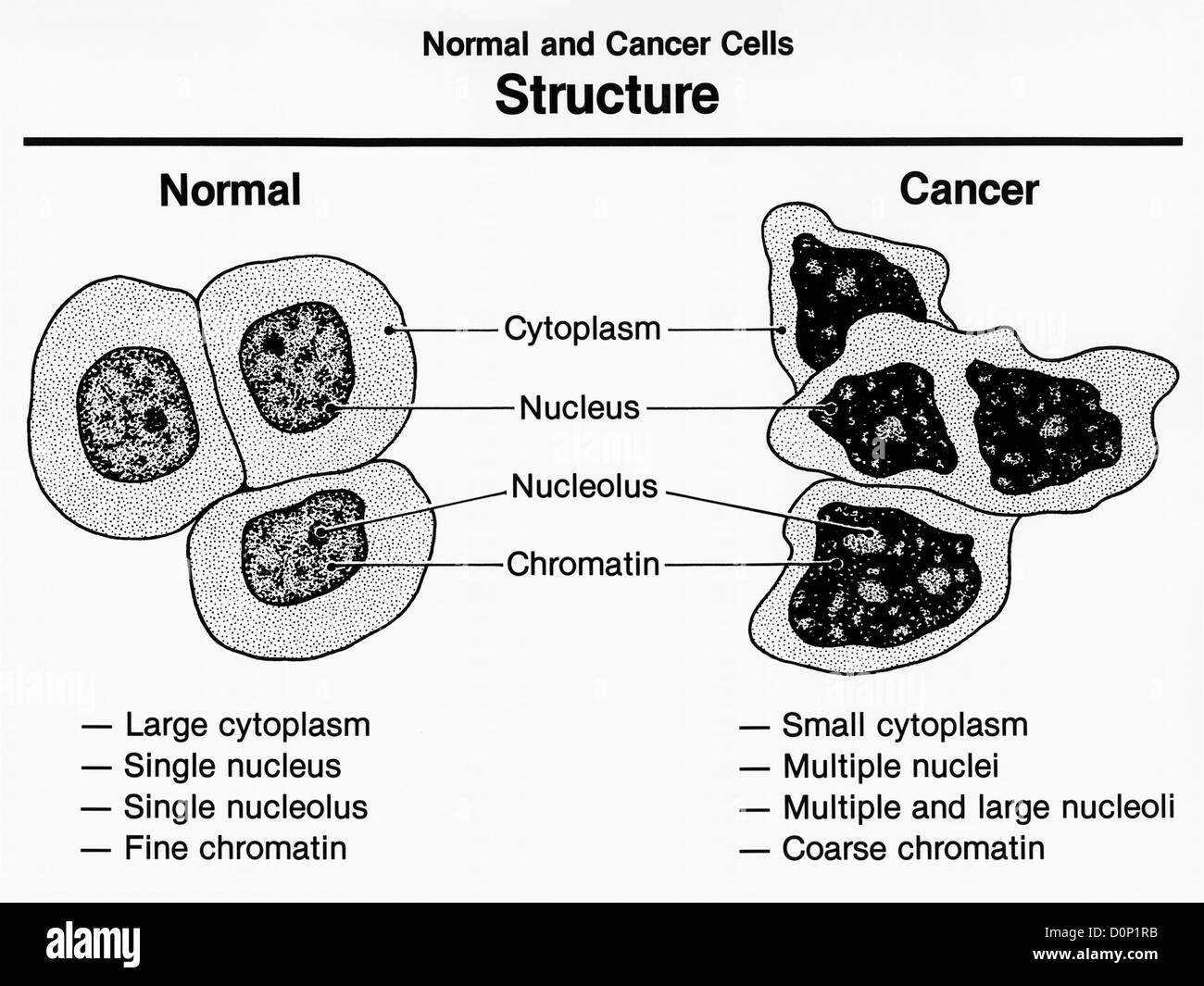
Cytologically, mitosis begins with prophase, when the nuclear envelope breaks down, the chromatin condenses to typical individualized rod-shaped chromosomes and the microtubule-based spindle apparatus starts assembling at the centrosomes (reviewed in [12]).During prometaphase and metaphase, the chromosomes are captured and concentrated in the middle of the cell, the so-called metaphase plate
Mitosis in Cancer Cells vs Regular Cells: Prohpase to Telophase Biology Diagrams
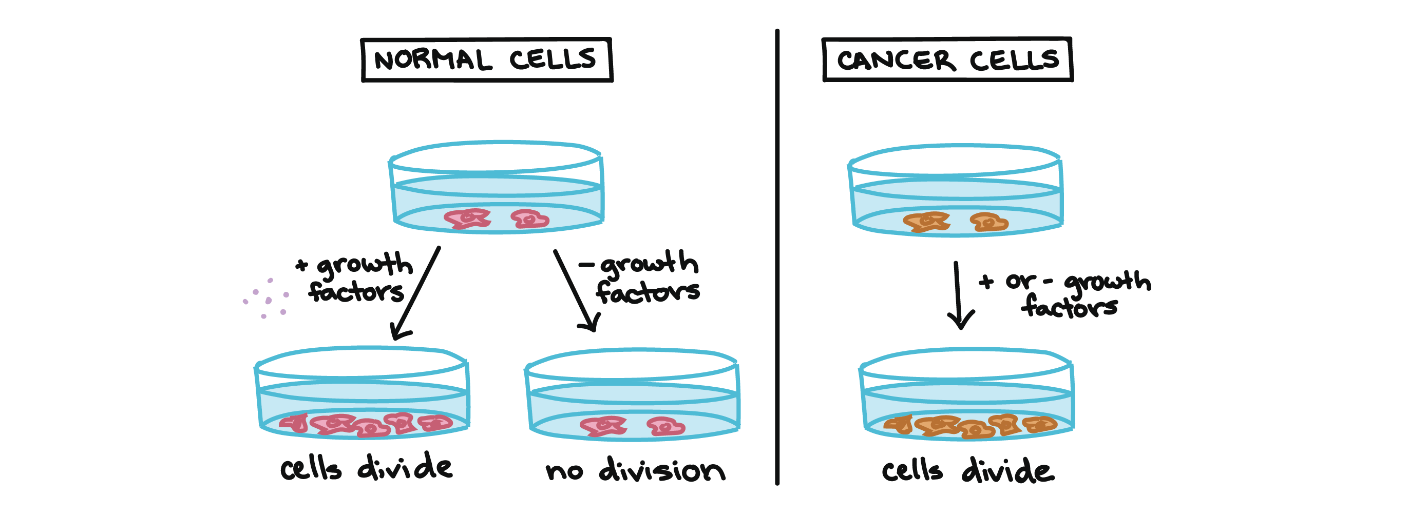
The challenge is to identify cell-cycle regulators that are essential for the mitosis of cancer cells rather than normal cells. The discovery of numerous mitotic kinases that are often overexpressed in cancers started a race to develop an arsenal of anti-mitotic drugs (Jackson et al., 2007; Lapenna and Giordano, 2009; Malumbres, 2011).
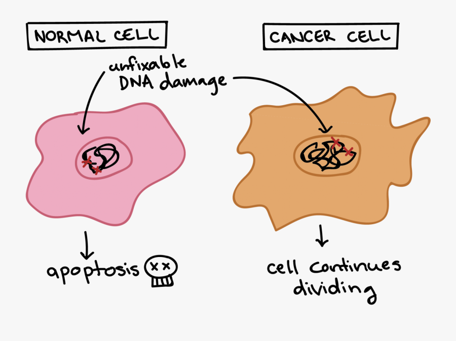
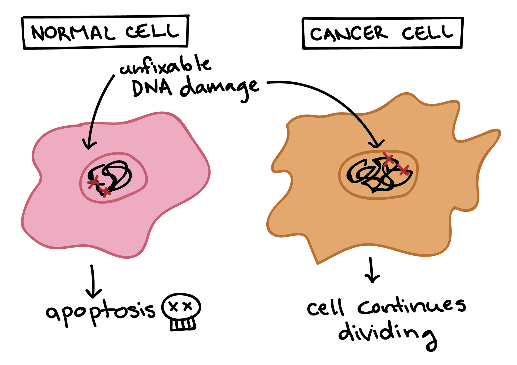
However, during this cell cycle, there are many situations where mistakes are made by the cycle or by a regulating system that causes the cell to proliferate uncontrollably, leading to cancer. Somatic cells are the best know for this cell cycle and go through 2 main phases called interphase and mitosis. Any errors during mitosis can have significant consequences for cellular function and organismal health. These errors may include improper chromosome segregation or mutations in DNA that can lead to abnormal cell behavior. The Link Between Mitosis and Cancer. Cancer arises when normal cellular processes become dysregulated. Another example of a cell cycle protein targeted by drug therapy is WEE1. It's part of a quality-control mechanism that pauses the cycle between the G2 and M stages so genetic errors can be fixed before the cell divides. Cancer cells without a functioning p53 protein may have accumulated substantial DNA damage by the time they reach the M stage.
cancer therapies: where they stand Biology Diagrams
Mitosis in Cancer Cells Cheyenne Friedersdorff Department of Biology and Wildlife, University of Alaska BIOL 111X: Human Anatomy and Physiology Professor Don Larson June 23, 2023. The cell cycle is an interesting subject to broach. The different stages of mitosis and how the cell reproduces and forms new cells has caught my attention in class.
In conclusion, the cancer cells in mitosis resist to immune cells attack. This phenomena is highly dependent on HLA-G expression during cell division. These results suggest the use of proliferation index (Ki-67), which is directly related to mitotic index, as a marker of immune resistance. In addition, the use of mitotic inhibitors may result Checkpoint kinases 1 and 2 (Chk1 and 2) are DNA damage checkpoint proteins regulating p53 and Cdc25 to mediate cell-cycle arrest/apoptosis and Cdk1 activation, respectively. 15 As G2 checkpoints, they are important for ensuring cells with cellular damage are prevented from progressing into mitosis until the damage is repaired. Studies have shown that the inhibition of checkpoint genes promote

How Is Mitosis Related To Cancer? Biology Diagrams
Supersession of the mitotic checkpoints in cancer cells causes runaway damage to the cell's organelles, which are units within the cells that carry out specific functions. During normal mitosis, damaged organelles have a chance to repair and recover between cell divisions, but they don't have this opportunity when cell division doesn't stop.