
Femoral Artery Anatomy Diagram Biology Diagrams This human anatomy diagram with labels depicts and explains the details and or parts of the Femoral Artery Anatomy.Human anatomy diagrams and charts show internal organs, body systems, cells, conditions, sickness and symptoms information and/or tips to ensure one lives in good health. Branches of the Femoral Artery. This illustration shows the following structures: common femoral artery, deep femoral artery (femoral profunda), superficial femoral artery, perforating arteries, lateral circumflex artery, medial circumflex artery, descending branch of the lateral circumflex artery, anastomotica magna, and superior external and internal articular branches of the popliteal artery.
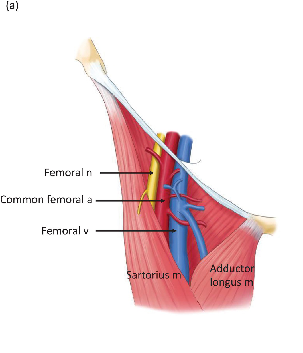
The superficial femoral artery flexes to follow the femur inferiorly and medially. At its distal end, it flexes again and descends posterior to the femur before forming the popliteal artery of the posterior knee and continuing on into the lower leg and foot. Several smaller arteries branch off from the superficial femoral artery to provide How To Do Femoral Vein Cannulation (Without Ultrasound Guidance) How To Do Femoral Vein Cannulation, Ultrasound-Guided How To Do Femoral Artery Cannulation, Ultrasound-Guided The femoral artery is a continuation of the external iliac artery and constitutes the major blood supply to the lower limb. In the thigh, the femoral artery passes through the femoral triangle, a wedge-shaped depression formed by muscles in the upper thigh.The medial and lateral boundaries of this triangle are formed by the medial margin of adductor longus and the medial margin of sartorius

Radiology Reference Article - Radiopaedia.org Biology Diagrams
Terminology. The femoral artery is commonly known clinically as the common femoral artery (CFA) and superficial femoral artery (SFA).The common femoral artery is the portion of the femoral artery between the inguinal ligament and branching of profunda femoris, and the superficial femoral artery is the portion distal to the branching of profunda femoris to the adductor hiatus.

The femoral artery is a large artery in the thigh and the main arterial supply to the thigh and leg. Diagram at MSU Archived July 17, 2011, at the Wayback Machine; QuantaFlo vs ABI in Peripheral Arterial Disease This page was last edited on 16 The femoral artery is the primary artery supplying blood to the lower limb. It is a continuation of the external iliac artery, beginning just below the inguinal ligament and extending down the thigh. [4] Location. The femoral artery originates from the external iliac artery as it passes under the inguinal ligament and enters the femoral triangle in the upper thigh.

Femoral artery Biology Diagrams
The femoral artery runs to the lower thigh and ends behind the knee. At the knee, the femoral artery becomes the popliteal artery. What are the parts of your femoral artery? The anatomy of the femoral artery includes: Common femoral artery: This first part of the femoral artery is an extension of the external iliac artery in your pelvis. It In the Leg. The popliteal artery descends down the posterior thigh, giving rise to genicular branches that supply the knee joint. It moves through the popliteal fossa, exiting between the gastrocnemius and popliteus muscles. At the lower border of the popliteus, the popliteal artery terminates by dividing into the anterior tibial artery and the tibioperoneal trunk.
