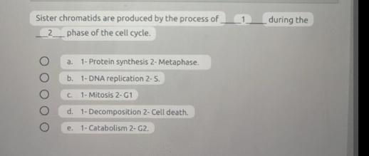
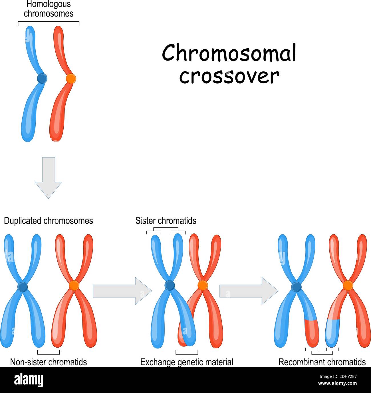
Figure 1 from Sister Chromatid Cohesion Control and Aneuploidy Biology Diagrams The paternal (blue) chromosome and the maternal (pink) chromosome are homologous chromosomes.Following chromosomal DNA replication, the blue chromosome is composed of two identical sister chromatids and the pink chromosome is composed of two identical sister chromatids.In mitosis, the sister chromatids separate into the daughter cells, but are now referred to as chromosomes (rather than While sister chromatids are exact copies of each other, non-sister chromatids come from homologous chromosomes. They code for the same genes, but are not genetically identical. domain contains a dynamic arrangement of proteins that are involved in mitotic checkpoints and regulators of chromosome behavior.

If this process were essential for sister chromatid separation, one would expect to find cdc5 mutants that block anaphase initiation. Consistent with this possibility, in Drosophila, there are polo mutants that arrest in metaphase with unseparated sister chromatids (Donaldson et al., 2001). Whether this metaphase arrest can be attributed to a
00439-1/asset/862038a5-5e9f-46ee-97f9-0815382ef277/main.assets/gr2.jpg)
Sister Chromatids - Definition, Functions and Structure Biology Diagrams
The identical copies of each chromosome are known as sister chromatids, and they are tightly associated together through G2 phase and early mitosis. During metaphase of mitosis, sister chromatids are associated with the mitotic spindle, aligned along the central axis of the cell, and one sister from each pair is associated with a separate

Sister chromatids, identical genetic copies of a chromosome, exhibit a fundamental behavior during cell division. These paired structures remain physically attached until a specific point in mitosis, known as anaphase. This separation is orchestrated by the spindle fibers, which are composed of microtubules that attach to structures called kinetochores on the centromeres of the chromosomes. Previously, differential labeling of sister chromatids and their quantitative analysis of nonoverlapping volume indicated that the resolution between the sisters can be detected as soon as cells initiate mitosis, and it proceeds hand in hand with chromatin compaction through prophase (Nagasaka et al., 2016).Being based on volumetric analyses, however, this study was inapplicable for analyzing
Linking Chromosome Duplication and Segregation via Sister Chromatid ... Biology Diagrams
Centromere behavior also differs. In Anaphase I, cohesin proteins keep sister chromatids together, protected by Shugoshin. In Anaphase II, this protection is lost, allowing separase to cleave cohesin. The centromeres split, enabling chromatids to move independently. Errors and Resulting Consequences