MRI Student orientation Biology Diagrams Magnetic Resonance Imaging (MRI) is a powerful diagnostic tool that provides detailed internal images of the human body. However, interpreting these images can be complex and requires a foundational understanding of anatomy, MRI technology, and image analysis.

e-Anatomy is a high-quality anatomy and imaging content atlas.It is the most complete reference of human anatomy available on the Web, iPad, iPhone and Android devices. Explore detailed anatomical views and multiple modalities (over 8,900 anatomic structures and more than 870,000 translated medical labels) with images in CT, MRI, radiographs, anatomical diagrams and nuclear images. Learn anatomy faster with our 3D human anatomy models. Over 400 models, by region, by structure and by system. TeachMe Anatomy. MRI; Areas. Scalp; Cranial Fossae; Pterygopalatine Fossa; Infratemporal Fossa; Mastoid Fossa; Bones. Skull; 3D Human Body; 3D Human Body. Complete Anatomy. Male Body; Female Body; Anatomy by Region. Thorax

Body MRI Approach: Guide for Common Indications Biology Diagrams
Normal chest x ray. Radiological anatomy is where your human anatomy knowledge meets clinical practice. It gathers several non-invasive methods for visualizing the inner body structures. The most frequently used imaging modalities are radiography (X-ray), computed tomography (CT) and magnetic resonance imaging (MRI).X-ray and CT require the use of ionizing radiation while MRI uses a magnetic Understanding the hypothalamus is crucial due to its central role in maintaining body homeostasis. This small but complex brain region influences numerous physiological processes, impacting overall health and well-being. Advancements in MRI technology have enhanced our ability to study the hypothalamus's intricate structure and functions.
Peter S. Liu, MD, Guest Editor Much like the central role of mathematics in science and engineering, anatomy forms a basic building block and universal language for the field of medicine. Magnetic resonance imaging (MRI) provides exquisite anatomic detail through its superior tissue contrast, flexible imaging planes, and tissue-characterization capabilities. The slide presentation demonstrates an approach to body MRI for common indications in the abdomen and pelvis, incorporating clinical information, knowledge of imaging patterns, and various scoring paradigms. Suggested Readings. Chung R, Garratt J, Remer EM, et

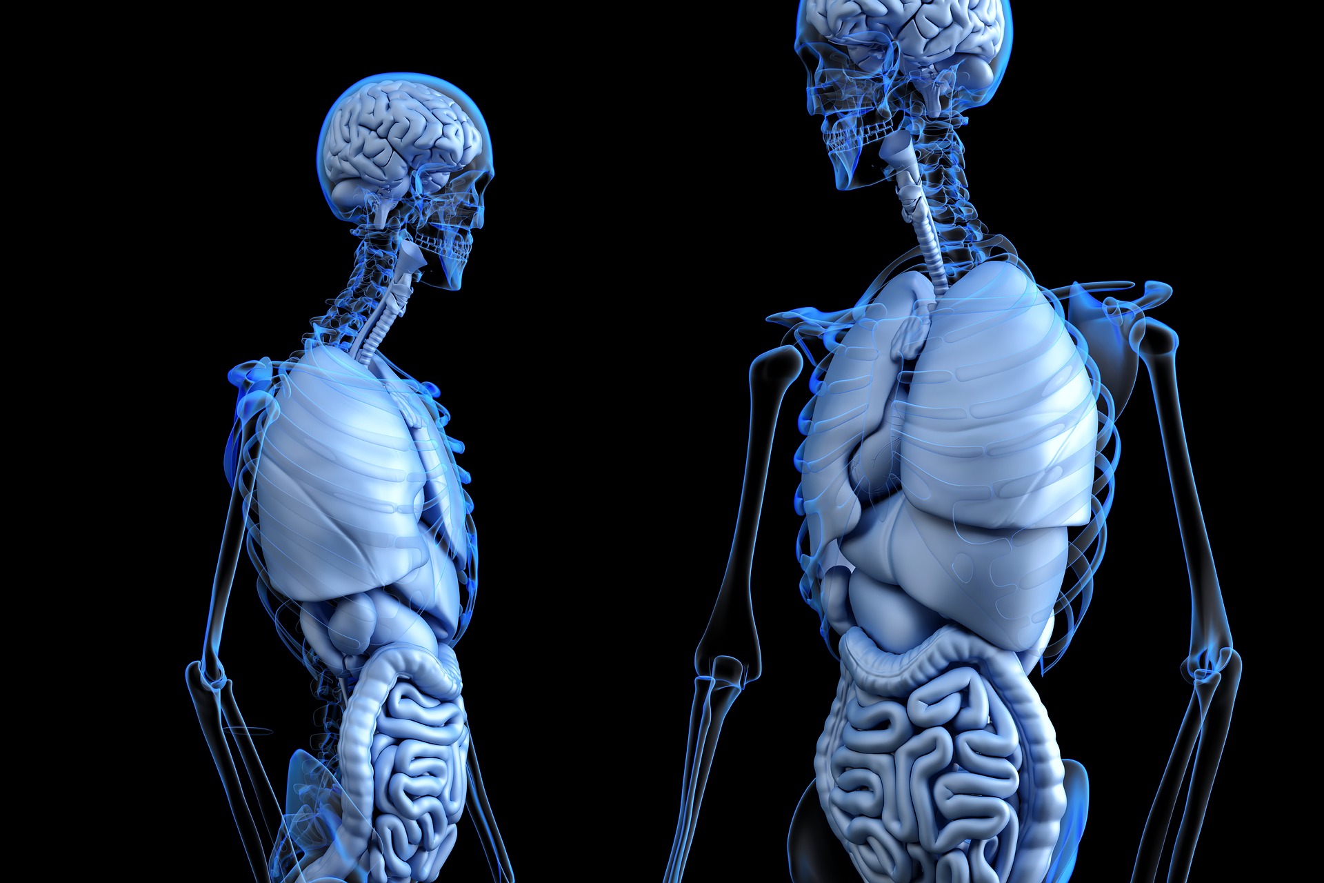
Anatomy, the Anatomy of Imaging Biology Diagrams
Visible Human Male. The Visible Human Male data set consists of MRI, CT, and anatomical images. Axial MRI images of the head and neck, and longitudinal sections of the rest of the body were obtained at 4mm intervals. The MRI images are 256 by 256 pixel resolution with each pixel made up of 12 bits of gray tone.

Contents: This atlas illustrates normal anatomy as evident on CT and MRI in multiple planes throughout the body. It includes most of the standard imaging planes used in diagnostic imaging and has detailed anatomic drawings to complement each image.