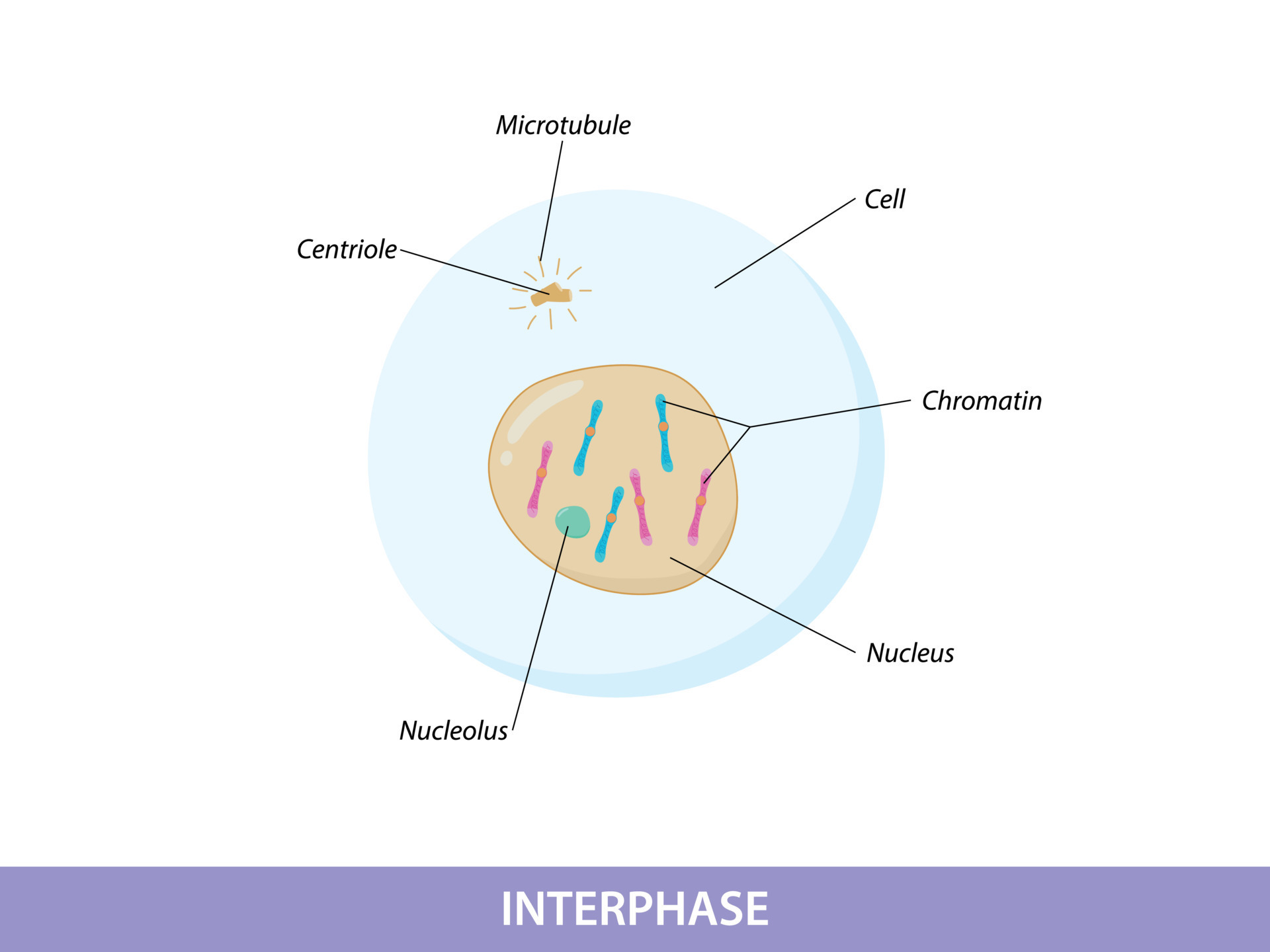
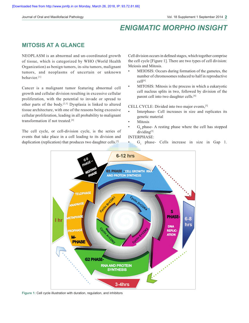
PDF Mitosis at a glance Biology Diagrams A normal resting cell exists in a state called interphase in which the chromatin is undifferentiated in the heavily-stained nucleus, as illustrated above. Before the cell enters the mitosis phase, it first undergoes a synthesis or S phase where each chromosome is duplicated and consists of two sister chromatids joined together by a specific DNA Mitosis is the phase of the cell cycle in which chromosomes in the nucleus are evenly divided between two cells. When the cell division process is complete, two daughter cells with identical genetic material are produced. Understanding the steps of mitosis is crucial in comprehending how organisms grow, develop, and repair tissues. The process of mitosis and its phases explained with steps in order. Learn its meaning, functions, & importance with examples & labeled picture
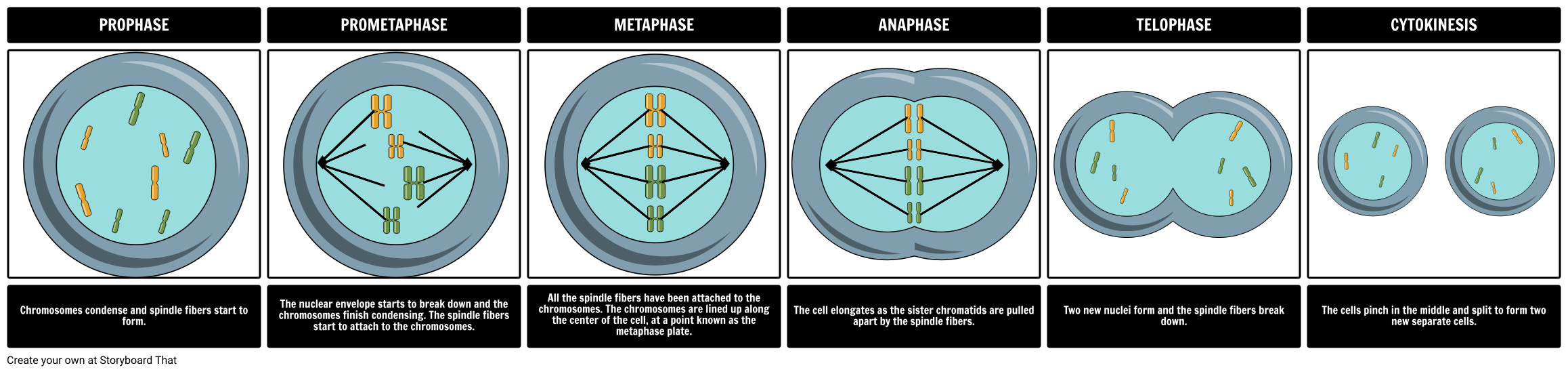
Curious about the stages of mitosis? Our complete guide goes deep on the 4 mitosis phases: prophase, metaphase, anaphase, and telophase. At the end of interphase, the cell has duplicated its chromosomes and is ready to move them into separate cells, called daughter cells. This occurs during the four steps of mitosis, called prophase, metaphase, anaphase and telophase. Check out what the mitosis phases look like under a microscope. Explore the stages of mitosis with detailed diagrams. Understand each phase and discover real-world applications of this essential cell division process.
Stages of Mitosis (with pictures) Flashcards Biology Diagrams
Learn about the stages of mitosis with a clear and detailed diagram. Discover the different phases of cell division, from prophase to telophase, and understand the important events that occur during each stage. This informative article provides a visual representation to help you understand the process of mitosis. Mitosis, the process by which a cell divides, consists of four distinct stages: prophase, metaphase, anaphase, and telophase. Drawing the stages of mitosis is an essential skill for students of biology, allowing them to visualize and understand the complex processes involved in cell division. The diagrams depict the changes in chromosome structure, spindle fiber formation, and nuclear membrane