SOLUTION Anatomy of oral cavity Biology Diagrams The oral cavity, or more commonly known as the mouth or buccal cavity, serves as the first portion of the digestive system. It consists of several different anatomically different aspects that work together effectively and efficiently to perform several functions. These aspects include the lips, tongue, palate, and teeth. Although a small compartment, the oral cavity is a unique and complex The borders of the oral cavity are the: cheeks or buccal surface (Latin, bucca, "cheek") - forming the sides; hard and soft palates - forming the "roof" tongue or lingual surface (Latin: lingua, "tongue") - forming the "floor."; There are four shapes of teeth. These shapes are the basis for the full names of the teeth:

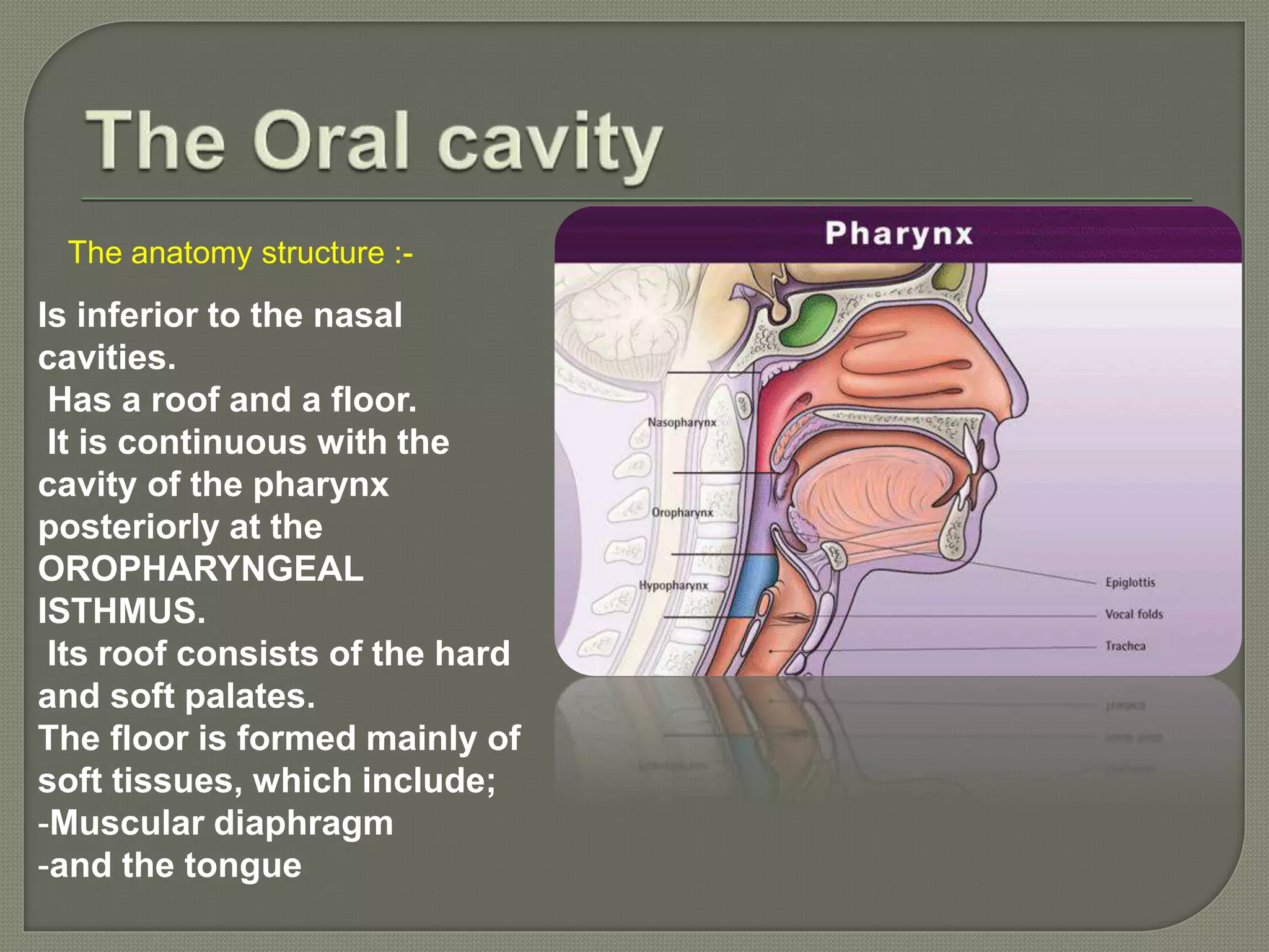
The human oral cavity refers to the mouth, and all its parts including the lips, tongue, teeth, and the roof and floor of the mouth [1, 2]. Oral Cavity Anatomy and Structure. The oral cavity is located just beneath the nasal cavity, the two being separated by the palate [4]. The oral cavity, better known as the mouth, is the start of the alimentary canal. It has three major functions: Digestion - receives food, preparing it for digestion in the stomach and small intestine.; Communication - modifies the sound produced in the larynx to create a range of sounds.; Breathing - acts as an air inlet in addition to the nasal cavity.
Oral Cavity Definition, Anatomy, Functions, Diagram Biology Diagrams
The oral cavity, commonly referred to as the mouth, is the initial part of the digestive system and plays an essential role in processes such as ingestion, speech, and breathing. Textbook of Oral Anatomy, Physiology, and Occlusion (3rd ed.). Elsevier. pp. 78-80. ISBN 978-8131248874. Oral cavity. The oral cavity is situated anteriorly on the face, under the nasal cavities.It is bounded by a roof, a floor and lateral walls. Anteriorly it opens to the face through the oral fissure, while posteriorly the oral cavity communicates with the oropharynx through a narrow passage called the oropharyngeal isthmus (also termed the isthmus of the fauces). This e-Anatomy module contains 114 illustrations on the oral cavity, the mouth, the tongue and the salivary glands. These fully annotated anatomical illustrations are presented as a comprehensive atlas of the oral cavity, specially designed for medical students, medicine residents and healthcare professionals. Material and methods
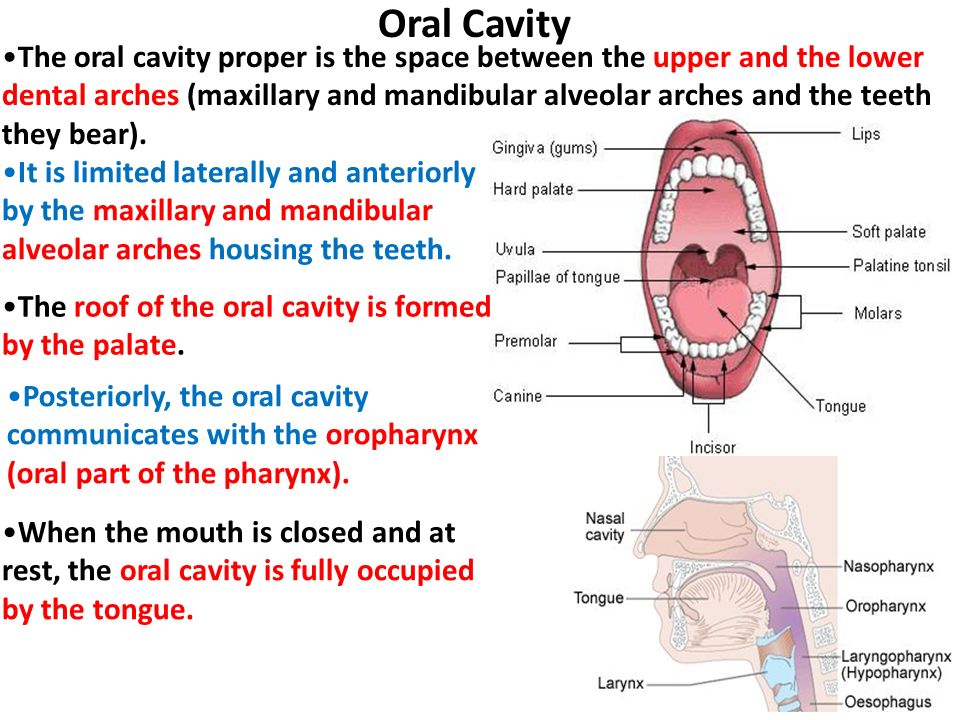
mouth, in human anatomy, orifice through which food and air enter the body. The mouth opens to the outside at the lips and empties into the throat at the rear; its boundaries are defined by the lips, cheeks, hard and soft palates, and glottis.It is divided into two sections: the vestibule, the area between the cheeks and the teeth, and the oral cavity proper.
Definition, Anatomy, & Function Biology Diagrams
Moving further inside is the oral cavity proper, bounded by the dental arches anteriorly and laterally, and the palate superiorly. The oral cavity proper is mainly occupied by the tongue. Now let's get back to that slice of pizza. To eat it, first you open your mouth, and the opening through which the food enters is called the oral fissure. Anatomy of the mouth. Floor of the mouth with lingual frenum and sublingual fold. The mouth consists of two regions: the vestibule and the oral cavity proper. The vestibule is the area between the teeth, lips and cheeks. [3] The oral cavity is bounded at the sides and in front by the alveolar process (containing the teeth) and at the back by the isthmus of the fauces.