SOLUTION Human Anatomy and Physiology Lab Exam Tissue Cell Diagrams Biology Diagrams Tissues are the foundation of the human body, consisting of groups of cells that work together to perform specific functions.[6] They form the basis for organs and systems, enabling the body to carry out complex tasks necessary for survival. Understanding the structure, types, and roles of tissues is critical in the study of anatomy and The amount and structure of each component correlate with the function of the tissue, from the rigid ground substance in bones supporting the body to the inclusion of specialized cells; for example, a phagocytic cell that engulfs pathogens and also rids tissue of cellular debris. Functions of Connective Tissues Tissues are groups of cells that have a similar structure and act together to perform a specific function. The word tissue comes from a form of an old French verb meaning "to weave". There are four different types of tissues in animals: connective, muscle, nervous, and epithelial. In plants, tissues are divided into three types: vascular, ground, and epidermal. Groups of tissues make up

Tissues provide the numerous functions of organs necessary to maintain biological life. This lab exercise seeks to introduce the various tissues found in the human body and to Cellular nature- Cells in epithelium fit closely together side by side and sometimes atop each other to form sheets of cells. These sheets are held together by

Tissue - Definition and Types of Tissues Biology Diagrams
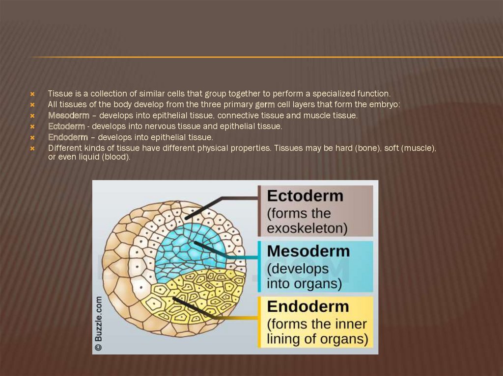
Tissue Membranes. A tissue membrane is a thin layer or sheet of cells that covers the outside of the body (for example, skin), the organs (for example, pericardium), internal passageways that lead to the exterior of the body (for example, abdominal mesenteries), and the lining of the moveable joint cavities.There are two basic types of tissue membranes: connective tissue and epithelial Figure 3.1.2 shows the types of tissues and organs associated with the each of the three germ layers. Note that epithelial tissue originates in all three layers, whereas nervous tissue derives primarily from the ectoderm and muscle tissue from mesoderm. Figure 3.1.2. Embryonic origin of tissues and major organs. Tissue Membranes

Cells, Tissues, & Membranes. This section provides detailed information about cell structure and function, four basic types of tissue in the human body, and the different types of membranes found in the body.

PDF Classification of Tissues Biology Diagrams
Tissue Membranes. A tissue membrane is a thin layer or sheet of cells that either covers the outside of the body (e.g., skin), lines an internal body cavity (e.g., peritoneal cavity), lines a vessel (e.g., blood vessel), or lines a movable joint cavity (e.g., synovial joint). Two basic types of tissue membranes are recognized based on the primary tissue type composing each: connective tissue